Denna Roshannon
  • Blog
  • About
  • Contact

Do Fallen Arches Need Surgery?

7/3/2017

 
Overview

Adult Acquired Flat Foot

A fallen arch is when an arch, even one that has always been flat, has gotten flatter. There are several reasons for this. The bottom of heel tilts toward the inside, making the inside of the ankle appear convex or bowed. The feet often appear to be rotates outward, like a duck. And the arch sags toward the ground. Fallen arches? are more than just flat feet. Having flat feet is not a disease. It is a description of the shape of the foot. Most flat feet have been like that all of the person?s life. Usually they are not painful.

Causes

Just as there are many different causes of flat feet, there are also many different treatment options. The most important aspect of treatment is determining the exact type or underlying cause of flat feet that you have. Foot and ankle specialists can determine this through thorough clinical examination and special imaging studies (e.g., x-rays, computed tomography, and/or magnetic resonance imaging). Conservative treatment is effective in the vast majority of flat foot cases, and consists of things such as insoles, splints, manipulation, or casting. Surgery is required much less frequently, and is reserved only for some of the severe types of flat foot that do not respond to conservative therapy. You may have noticed that one common element in the conservative treatment of all types of flat feet is orthoses. Many companies now manufacture semi-custom orthotic devices that not only improve comfort, but also seek to control abnormal motion of the foot. These over-the-counter inserts, in the $25 to $50 range, are an economical treatment that may help a majority of people. Unfortunately, these semi-custom devices will not fit everyone perfectly, and those of us who differ too much from the average may respond better to custom orthotic devices. Custom inserts are prescribed by your foot and ankle specialist and are made individually from either a physical or computerized impression of your feet. The only drawback of custom orthoses is their cost, ranging anywhere from $300 to $500. Many physicians recommend trying over-the-counter inserts first (and even keep them in stock) as they may save their patients large sums of money.

Symptoms

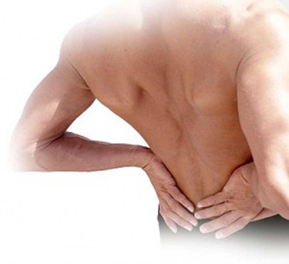
Fallen arches may induce pain in the heel, the inside of the arch, the ankle, and may even extend up the body into the leg (shin splints), knee, lower back and hip. You may also experience inflammation (swelling, redness, heat and pain) along the inside of the ankle (along the posterior tibial tendon). Additionally, you may notice some changes in the way your foot looks. Your ankle may begin to turn inward (pronate), causing the bottom of your heel to tilt outward. Other secondary symptoms may also show up as the condition progresses, such as hammertoes or bunions. You may also want to check your footprint after you step out of the shower. (It helps if you pretend you?re in a mystery novel, and you?re leaving wet, footprinty clues that will help crack the case.) Normally, you can see a clear imprint of the front of your foot (the ball and the toes) the heel, and the outside edge of your foot. There should be a gap (i.e. no footprinting) along the inside where your arches are. If your foot is flat, it?ll probably leave an imprint of the full bottom of your foot-no gap to be had. Your shoes may also be affected: because the ankle tilts somewhat with this condition, the heel of your shoes may become more worn on one side than another.

Diagnosis

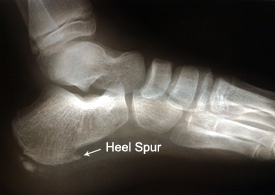
People who have flat feet without signs or symptoms that bother them do not generally have to see a doctor or podiatrist about them. However, if any of the following occur, you should see your GP or a podiatrist. The fallen arches (flat feet) have developed recently. You experience pain in your feet, ankles or lower limbs. Your unpleasant symptoms do not improve with supportive, well-fitted shoes. Either or both feet are becoming flatter. Your feet feel rigid (stiff). Your feet feel heavy and unwieldy. Most qualified health care professionals can diagnose flat feet just by watching the patient stand, walk and examining his/her feet. A doctor will also look at the patient's medical history. The feet will be observed from the front and back. The patient may be asked to stand on tip-toe while the doctor examines the shape and functioning of each foot. In some cases the physician may order an X-ray, CT (computed tomography) scan, or MRI (magnetic resonance imaging) scan.

pes planus treatment

Non Surgical Treatment

Orthotics. Interpod orthotics re-align and support the foot; therefore reducing any excessive stress when walking or during activity. Orthotics can assist with maintaining arch profile and allow for more effective functioning of joints. Footwear. A strong supportive, well fitted shoe may assist with reducing excessive pronation and support the joints of your feet. A supportive shoe will also help maximise the function of your Interpod orthotic. Padding may be applied to your shoes or feet by your practitioner to reduce excessive stress. Specific taping techniques can be applied by your practitioner to improve foot function. Your practitioner may advise certain stretches or exercises to assist with maintaining foot function and reduce painful symptoms. Pain medication such as NSAIDs (ibuprofen) may be advised by your practitioner. If all conservative options have been exhausted, then surgical correction of flat feet may be undertaken.

Surgical Treatment

Acquired Flat Feet

Surgery for flat feet is separated into three kinds: soft tissue procedures, bone cuts, and bone fusions. Depending on the severity of the flat foot, a person?s age, and whether or not the foot is stiff determines just how the foot can be fixed. In most cases a combination of procedures are performed. With flexible flat feet, surgery is geared at maintaining the motion of the foot and recreating the arch. Commonly this may involve tendon repairs along the inside of the foot to reinforce the main tendon that lifts the arch. When the bone collapse is significant, bone procedures are included to physically rebuild the arch, and realign the heel. The presence of bunions with flat feet is often contributing to the collapse and in most situations requires correction. With rigid flat feet, surgery is focused on restoring the shape of the foot through procedures that eliminate motion. In this case, motion does not exist pre-operatively, so realigning the foot is of utmost importance. The exception, are rigid flat feet due to tarsal coalition (fused segment of bone) in the back of the foot where freeing the blockage can restore function.

After Care

Time off work depends on the type of work as well as the surgical procedures performed. . A patient will be required to be non-weight bearing in a cast or splint and use crutches for four to twelve weeks. Usually a patient can return to work in one to two weeks if they are able to work while seated. If a person's job requires standing and walking, return to work may take several weeks. Complete recovery may take six months to a full year. Complications can occur as with all surgeries, but are minimized by strictly following your surgeon's post-operative instructions. The main complications include infection, bone that is slow to heal or does not heal, progression or reoccurrence of deformity, a stiff foot, and the need for further surgery. Many of the above complications can be avoided by only putting weight on the operative foot when allowed by your surgeon.

Do You Really Understand Heel Soreness?

7/1/2017

 
Overview

Painful Heel

Heel pain is most often caused by plantar fasciitis, a condition that is sometimes also called heel spur syndrome when a spur is present. Heel pain may also be due to other causes, such as a stress fracture, tendonitis, arthritis, nerve irritation, or, rarely, a cyst. Because there are several potential causes, it is important to have heel pain properly diagnosed. A foot and ankle surgeon is able to distinguish between all the possibilities and determine the underlying source of your heel pain.What Is Plantar Fasciitis? Plantar fasciitis is an inflammation of the band of tissue (the plantar fascia) that extends from the heel to the toes. In this condition, the fascia first becomes irritated and then inflamed, resulting in heel pain.

Causes

Heel pain is most often the result of overuse. Rarely, it may be caused by an injury. Your heel may become tender or swollen from shoes with poor support or shock absorption, running on hard surfaces, like concrete, running too often, tightness in your calf muscle or the Achilles tendon. Sudden inward or outward turning of your heel, landing hard or awkwardly on the heel. Conditions that may cause heel pain include when the tendon that connects the back of your leg to your heel becomes swollen and painful near the bottom of the foot, swelling of the fluid-filled sac (bursa) at the back of the heel bone under the Achilles tendon (bursitis). Bone spurs in the heel. Swelling of the thick band of tissue on the bottom of your foot (plantar fasciitis). Fracture of the heel bone that is related to landing very hard on your heel from a fall (calcaneus fracture).

Symptoms

The most common complaint is pain and stiffness in the bottom of the heel. Heel pain may be sharp or dull, and it may develop slowly over time or suddenly after intense activity. The pain is typically worse in the morning, when taking your first steps of the day. After standing or sitting for a while. When climbing stairs.

Diagnosis

Your doctor will perform a physical exam and ask questions about your medical history and symptoms, such as have you had this type of heel pain before? When did your pain begin? Do you have pain upon your first steps in the morning or after your first steps after rest? Is the pain dull and aching or sharp and stabbing? Is it worse after exercise? Is it worse when standing? Did you fall or twist your ankle recently? Are you a runner? If so, how far and how often do you run? Do you walk or stand for long periods of time? What kind of shoes do you wear? Do you have any other symptoms? Your doctor may order a foot x-ray. You may need to see a physical therapist to learn exercises to stretch and strengthen your foot. Your doctor may recommend a night splint to help stretch your foot. Surgery may be recommended in some cases.

Non Surgical Treatment

There are a number of treatments that can help relieve heel pain and speed up your recovery. These include resting your heel, try to avoid walking long distances and standing for long periods, regular stretching, stretching your calf muscles and plantar fascia, pain relief, using an icepack on the affected heel and taking painkillers, such as non-steroidal anti-inflammatory drugs (NSAIDs), wearing good-fitting shoes that support and cushion your foot, running shoes are particularly useful, using supportive devices such as orthoses (rigid supports that are put inside the shoe) or strapping. Around four out of five cases of heel pain resolve within a year. However, having heel pain for this length of time can often be frustrating and painful. In around one in 20 cases, the above treatments are not enough, and surgery may be recommended to release the plantar fascia.

Surgical Treatment

With the advancements in technology and treatments, if you do need to have surgery for the heel, it is very minimal incision that?s done. And the nice thing is your recovery period is short and you should be able to bear weight right after the surgery. This means you can get back to your weekly routine in just a few weeks. Recovery is a lot different than it used to be and a lot of it is because of doing a minimal incision and decreasing trauma to soft tissues, as well as even the bone. So if you need surgery, then your recovery period is pretty quick.

heel pad anatomy

Prevention

Heel Pain

You should always wear footwear that is appropriate for your environment and day-to-day activities. Wearing high heels when you go out in the evening is unlikely to be harmful. However, wearing them all week at work may damage your feet, particularly if your job involves a lot of walking or standing. Ideally, you should wear shoes with laces and a low to moderate heel that supports and cushions your arches and heels. Avoid wearing shoes with no heels. Do not walk barefoot on hard ground, particularly while on holiday. Many cases of heel pain occur when a person protects their feet for 50 weeks of the year and then suddenly walks barefoot while on holiday. Their feet are not accustomed to the extra pressure, which causes heel pain. If you do a physical activity, such as running or another form of exercise that places additional strain on your feet, you should replace your sports shoes regularly. Most experts recommend that sports shoes should be replaced after you have done about 500 miles in them. It is also a good idea to always stretch after exercising, and to make strength and flexibility training a part of your regular exercise routine.

Leg Length Discrepancy Testing

6/29/2017

 
Overview

Surgery is another option. In some cases the longer extremity can be shortened, but a major shortening may weaken the muscles of the extremity. In growing children, lower extremities can also be equalized by a surgical procedure that stops the growth at one or two sites of the longer extremity, while leaving the remaining growth undisturbed. Your physician can tell you how much equalization can be attained by surgically halting one or more growth centers. The procedure is performed under X-ray control through very small incisions in the knee area. This procedure will not cause an immediate correction in length. Instead, the LLD will gradually decrease as the opposite extremity continues to grow and "catch up." Timing of the procedure is critical; the goal is to attain equal length of the extremities at skeletal maturity, usually in the mid- to late teens. Disadvantages of this option include the possibility of slight over-correction or under-correction of the LLD and the patient?s adult height will be less than if the shorter extremity had been lengthened. Correction of significant LLDs by this method may make a patient?s body look slightly disproportionate because of the shorter legs.Leg Length Discrepancy

Causes

Sometimes the cause of LLD is unknown, yet the pattern or combination of conditions is consistent with a certain abnormality. Examples include underdevelopment of the inner or outer side of the leg (hemimelias) or (partial) inhibition of growth of one side of the body of unknown cause (hemihypertrophy). These conditions are present at birth, but the limb length difference may be too small to be detected. As the child grows, the LLD increases and becomes more noticeable. In hemimelia, one of the two bones between the knee and the ankle (tibia or fibula) is abnormally short. There also may be associated foot or knee abnormalities. Hemihypertrophy or hemiatrophy are rare conditions in which there is a difference in length of both the arm and leg on only one side of the body. There may also be a difference between the two sides of the face. Sometimes no cause can be found. This type of limb length is called idiopathic. While there is a cause, it cannot be determined using currect diagnostic methods.

Symptoms

The symptoms of limb deformity can range from a mild difference in the appearance of a leg or arm to major loss of function of the use of an extremity. For instance, you may notice that your child has a significant limp. If there is deformity in the extremity, the patient may develop arthritis as he or she gets older, especially if the lower extremities are involved. Patients often present due to the appearance of the extremity (it looks different from the other side).

Diagnosis

A doctor will generally take a detailed medical history of both the patient and family, including asking about recent injuries or illnesses. He or she will carefully examine the patient, observing how he or she moves and stands. If necessary, an orthopedic surgeon will order X-ray, bone age determinations and computed tomography (CT) scans or magnetic resonance imaging (MRI).

Non Surgical Treatment

You and your physician should discuss whether treatment is necessary. For minor LLDs in adults with no deformity, treatment may not be necessary. Because the risks may outweigh the benefits, surgical treatment to equalize leg lengths is usually not recommended if the difference is less than one inch. For these small differences, your physician may recommend a shoe lift. A lift fitted to the shoe can often improve your walking and running, as well as relieve back pain caused by LLD. Shoe lifts are inexpensive and can be removed if they are not effective. They do, however, add weight and stiffness to the shoe.

Leg Length

how to increase height in 1 week

Surgical Treatment

Lengthening is usually done by corticotomy and gradual distraction. This technique can result in lengthenings of 25% or more, but typically lengthening of 15%, or about 6 cm, is recommended. The limits of lengthening depend on patient tolerance, bony consolidation, maintenance of range of motion, and stability of the joints above and below the lengthened limb. Numerous fixation devices are available, such as the ring fixator with fine wires, monolateral fixator with half pins, or a hybrid frame. The choice of fixation device depends on the desired goal. A monolateral device is easier to apply and better tolerated by the patient. The disadvantages of monolateral fixation devices include the limitation of the degree of angular correction that can concurrently be obtained; the cantilever effect on the pins, which may result in angular deformity, especially when lengthening the femur in large patients; and the difficulty in making adjustments without placing new pins. Monolateral fixators appear to have a similar success rate as circular fixators, especially with more modest lengthenings (20%).

Leg Length Discrepancy Symptoms Running

6/26/2017

 
Overview

Approximately 75% of us present with one leg longer than the other. It?s staggering, literally, that so many people walk about with an imbalance. Yet to have one leg longer than the other doesn?t seem to create pain for everyone but for those that it does it brings pain in a myriad of dysfunction from TMJ, headaches, low back pain, IBS, bladder problems, sexual dysfunction, sacroiliac joint pain, pubis dysfunction, groin strain, gluteal dysfunction as well as the formation of trigger points.Leg Length Discrepancy

Causes

From an anatomical stand point, the LLD could have been from hereditary, broken bones, diseases and joint replacements. Functional LLD can be from over pronating, knee deformities, tight calves and hamstrings, weak IT band, curvature in the spine and many other such muscular/skeletal issues.

Symptoms

If your child has one leg that is longer than the other, you may notice that he or she bends one leg. Stands on the toes of the shorter leg. Limps. The shorter leg has to be pushed upward, leading to an exaggerated up and down motion during walking. Tires easily. It takes more energy to walk with a discrepancy.

Diagnosis

A doctor will generally take a detailed medical history of both the patient and family, including asking about recent injuries or illnesses. He or she will carefully examine the patient, observing how he or she moves and stands. If necessary, an orthopedic surgeon will order X-ray, bone age determinations and computed tomography (CT) scans or magnetic resonance imaging (MRI).

Non Surgical Treatment

For minor limb length discrepancy in patients with no deformity, treatment may not be necessary. Because the risks may outweigh the benefits, surgical treatment to equalize leg lengths is usually not recommended if the difference is less than 1 inch. For these small differences, the physician may recommend a shoe lift. A lift fitted to the shoe can often improve walking and running, as well as relieve any back pain that may be caused by the limb length discrepancy. Shoe lifts are inexpensive and can be removed if they are not effective.

Leg Length Discrepancy Insoles

can gym help in increasing height?

Surgical Treatment

Surgery to shorten the longer leg. This is less involved than lengthening the shorter leg. Shortening may be done in one of two ways. Closing the growth plate of the long leg 2-3 years before growth ends (around age 11-13), letting the short leg catch up. This procedure is called an epiphysiodesis. Taking some bone from the longer leg once growth is complete to even out leg lengths. Surgery to lengthen the shorter leg. This surgery is more involved than surgery to shorten a leg. During this surgery, cuts are made in the leg bone. An external metal frame and bar are attached to the leg bone. This frame and bar slowly pull on the leg bone, lengthening it. The frame and bar must be worn constantly for months to years. When the frame and bar are removed, a leg cast is required for several months. This surgery requires careful and continued follow-up with the surgeon to be sure that healing is going well.

Mortons Neuroma Symptoms

5/31/2017

 
Overview

interdigital neuromaA Morton's neuroma usually develops between the third and fourth toes. Less commonly, it develops between the second and third toes. Other locations are rare. It also is rare for a Morton's neuroma to develop in both feet at the same time. The condition is much more common in women than men, probably as a result of wearing high-heeled, narrow-toed shoes. This style of shoe tends to shift the bones of the feet into an abnormal position, which increases the risk that a neuroma will form. Being overweight also increases the risk of a Morton's neuroma.

Causes

Various factors have been implicated in the precipitation of Morton's neuroma. Morton's neuroma is known to develop as a result of chronic nerve stress and irritation, particularly with excessive toe dorsiflexion. Poorly fitting and constricting shoes (ie, small toe box) or shoes with heel lifts often contribute to Morton's neuroma. Women who wear high-heeled shoes for a number of years or men who are required to wear constrictive shoe gear are at risk. A biomechanical theory of causation involves the mechanics of the foot and ankle. For instance, individuals with tight gastrocnemius-soleus muscles or who excessively pronate the foot may compensate by dorsiflexion of the metatarsals subsequently irritating of the interdigital nerve. Certain activities carry increased risk of excessive toe dorsiflexion, such as prolonged walking, running, squatting, and demi-pointe position in ballet.

Symptoms

Pain is usually increased by forefoot weight bearing activities (such as running), with narrow-fitting footwear, or with high heeled shoes. It is usually painful to firmly touch the affected region and, in chronic cases, pain and sometimes an audible click, may be heard when squeezing the foot and toes together with the hand. Often a localized area of swelling may be evident at the site of injury.

Diagnosis

Metatarsal bones will be examined clinically, and often an x-ray will be taken to assess the particular case and ensure against other conditions, including fracture. When the foot is examined by a doctor, he may feel a characteristic ?click,? referred to as Mulder?s sign, and the interspaces between toe bones will often be tender. The doctor may put pressure on these areas to localize the site of pain and test for other conditions, including calluses or stress fractures. Range of motion tests will also be applied to rule out arthritis or joint inflammations. X-rays may be required to ensure there are no stress fractures or arthritis within the joints that join the toes to the foot. Tenderness in one or more metatarsal bones may imply a pre-stress fracture or stress-fracture. An ultrasound scan may be used to confirm diagnosis of Morton?s Neuroma, as x-ray will not detect the condition, (but can confirm that the bones are uninjured).

Non Surgical Treatment

Sclerosing alcohol injections are an increasingly available treatment alternative if the above management approaches fail. Dilute alcohol (4%) is injected directly into the area of the neuroma, causing toxicity to the fibrous nerve tissue. Frequently, treatment must be performed 2-4 times, with 1-3 weeks between interventions. An 60-80% success rate has been achieved in clinical studies, equal to or exceeding the success rate for surgical neurectomy with fewer risks and less significant recovery. If done with more concentrated alcohol under ultrasound guidance, the success rate is considerably higher and fewer repeat procedures are needed. Radio Frequency Ablation is also used in the treatment of Morton's Neuroma The outcomes appear to be equally or more reliable than alcohol injections especially if the procedure is done under ultrasound guidance.plantar neuroma

Surgical Treatment

About one person in four will not require any surgery for Morton's neuroma and their symptoms can be controlled with footwear modification and steroid/local anaesthetic injections. Of those who choose to have surgery, about three out of four will have good results with relief of their symptoms. Recurrent or persisting (chronic) symptoms can occur after surgery. Sometimes, decompression of the nerve may have been incomplete or the nerve may just remain 'irritable'. In those who have had cutting out (resection) of the nerve (neurectomy), a recurrent or 'stump' neuroma may develop in any nerve tissue that was left behind. This can sometimes be more painful than the original condition.

How Shoe Lifts Cure Leg Length Difference

2/20/2016

 
There are actually not one but two different kinds of leg length discrepancies, congenital and acquired. Congenital indicates you are born with it. One leg is structurally shorter in comparison to the other. Through developmental stages of aging, the human brain senses the walking pattern and identifies some variation. Our bodies usually adapts by tilting one shoulder over to the "short" side. A difference of under a quarter inch is not grossly abnormal, does not need Shoe Lifts to compensate and commonly does not have a profound effect over a lifetime.

Shoe Lifts

Leg length inequality goes largely undiagnosed on a daily basis, however this issue is simply solved, and can reduce numerous instances of chronic back pain.

Treatment for leg length inequality typically consists of Shoe Lifts. These are typically affordable, commonly being under twenty dollars, compared to a custom orthotic of $200 or even more. Differences over a quarter inch can take their toll on the spine and should probably be compensated for with a heel lift. In some cases, the shortage can be so extreme that it requires a full lift to both the heel and sole of the shoe.

Chronic back pain is easily the most widespread health problem afflicting men and women today. Over 80 million people suffer from back pain at some point in their life. It is a problem which costs employers huge amounts of money yearly due to lost time and production. Innovative and better treatment solutions are always sought after in the hope of reducing the economic impact this issue causes.

Shoe Lifts

Men and women from all corners of the world suffer from foot ache as a result of leg length discrepancy. In most of these situations Shoe Lifts can be of very helpful. The lifts are capable of decreasing any discomfort and pain in the feet. Shoe Lifts are recommended by countless qualified orthopaedic physicians.

In order to support the body in a healthy and balanced fashion, the feet have got a critical job to play. Inspite of that, it is sometimes the most overlooked region of the human body. Many people have flat-feet meaning there is unequal force placed on the feet. This causes other parts of the body such as knees, ankles and backs to be impacted too. Shoe Lifts make sure that suitable posture and balance are restored.

What Are The Leading Causes Of Posterior Calcaneal Spur

9/27/2015

 
Heel Spur

Overview

Heel spurs are tiny protruding calcium deposits that can develop near the base of your heel bone. They can be caused by repetitive activities, such as dancing or running, or they can form in association with plantar fasciitis, which is an inflammation of the ligament (plantar fascia) on the bottom of your foot. When the plantar fascia is tight and pulls on your heel bone, the bone releases calcium to try to heal itself. The excess deposits of calcium can sometimes form heel spurs.

Causes

At the onset of this condition, pain and swelling become present, with discomfort particularly noted as pushing off with the toes occurs during walking. This movement of the foot stretches the fascia that is already irritated and inflamed. If this condition is allowed to continue, pain is noticed around the heel region because of the newly formed bone, in response to the stress. This results in the development of the heel spur. It is common among athletes and others who run and jump a significant amount.

Heel Spur

Symptoms

It is important to be aware that heel spurs may or may not cause symptoms. Symptoms are usually related to the plantar fasciitis. You may experience significant pain and it may be worse in the morning when you first wake up or during certain physical activities such as, walking, jogging, or running.

Diagnosis

Sharp pain localized to the heel may be all a doctor needs to understand in order to diagnose the presence of heel spurs. However, you may also be sent to a radiologist for X-rays to confirm the presence of heel spurs.

Non Surgical Treatment

Ice compresses, stretching exercises, night splint for traction of the leg muscles to stretch the muscle in the back of the leg, and massage of the back of the leg, along with padding and heel cushions are also things that you can do at home. The number one recommendation for relief of heel pain is wearing good shoe gear. Good shoe gear usually consists of a sturdy, solid shoe. Heel pain is not relieved by a soft, ill supported shoe. Shoes such as Nike, K-Swiss, and Avia are the best shoes for this condition. Custom orthotics are highly recommended. Physical therapy is another way physicians treat this condition. Ice packs, muscle stimulation, ultra sound, paraffin baths, and the new Plantar Fascitis Night Splint are also helpful. If all these conservative measures fail to relieve the pain, then surgery is indicated. The newer minimal incision surgeries such as the Endoscopic plantar fasciotomy surgery is extremely beneficial for this condition, and for earlier ambulation, the use of the newer Cast Walking Boot is recommended.

Surgical Treatment

Usually, heel spurs are curable with conservative treatment. If not, heel spurs are curable with surgery, although there is the possibility of them growing back. About 10% of those who continue to see a physician for plantar fascitis have it for more than a year. If there is limited success after approximately one year of conservative treatment, patients are often advised to have surgery.

What Are The Symptoms Of Inferior Calcaneal Spur

9/22/2015

 
Inferior Calcaneal Spur

Overview

A heel spur also known as a calcaneal spur, is a pointed bony outgrowth of the heel bone (calcaneus). Heel spurs do not always cause pain and often are discovered incidentally on X-rays taken for other problems. Heel spurs can occur at the back of the heel and also under the heel bone on the sole of the foot, where they may be associated with the painful foot condition plantar fasciitis.

Causes

Bone spurs can occur all over the body including the spine, shoulders, hands, hips and feet. The feet are a common place to find them. A heel spur happens when the body tries to mend itself. Building extra bone is one way your body tries to correct a weakness. Wearing shoes that are too tight in the heel can cause bone spurs. More women than men get heel spurs because of the kinds of shoes they wear. Athletes who stress their feet and legs routinely are also prone to heel spurs. Being overweight can also indirectly cause heel spurs by over-exerting the plantar fascia. Some heel spurs are caused by the aging process, in which the cartilage covering the ends of bones wears away. This process can lead to pain, swelling and spur formation. Stress-related problems with the plantar fascia frequently lead to heel spurs.

Calcaneal Spur

Symptoms

Heel spurs result in a jabbing or aching sensation on or under the heel bone. The pain is often worst when you first arise in the morning and get to your feet. You may also experience pain when standing up after prolonged periods of sitting, such as work sessions at a desk or car rides. The discomfort may lessen after you spend several minutes walking, only to return later. Heel spurs can cause intermittent or chronic pain.

Diagnosis

A heel spur is often seen on X-ray as a bony protrusion, which can vary in size. However, because a Heel Spur only indicates increased load on the plantar fascia, and not pain, an ultra sound may be required to assess other actual cause of the heel pain such and may include checking to see if the plantar fascia is inflamed or degenerated.

Non Surgical Treatment

Many treatment options exist, and good results are often observed. Generally, a calcaneal spur develops when proper care is not given to the foot and heels. It is often seen as a repetitive stress injury, and thus lifestyle modification is typically the basic course of management strategies. To alleviate heel spur pain, a person should begin doing foot and calf workouts. Strong muscles in the calves and lower legs will help take the stress off the bone and thus help cure or prevent heel spurs. Icing the area is an effective way to get immediate pain relief.

Surgical Treatment

When chronic heel pain fails to respond to conservative treatment, surgical treatment may be necessary. Heel surgery can provide pain relief and restore mobility. The type of procedure used is based on examination and usually consists of releasing the excessive tightness of the plantar fascia, called a plantar fascia release. The procedure may also include removal of heel spurs.

Bursitis Of The Heel Warning Signs

8/25/2015

 
Overview

Retrocalcaneal bursitis most commonly occurs as s result of repetitive activity that encourages the calf muscles to tighten and shorten from overuse, like repetitively wearing high heels, running and even wearing tight shoes that pinch at the back of the heel. Symptoms normally include a constant dull ache or burning pain at the back of the heel that is aggravated by any touch or pressure from tight shoes or movement of the ankle joint. There will normally be noticeable swelling around the back of the heel. In cases of bursitis caused by infection the skin around the affected joint will appear red and will feel incredibly warm to the touch. Additional symptoms are a high temperature and feverish chills. Retrocalcaneal bursitis is very similar to Achilles bursitis as the bursae are very close in proximity and symptoms are almost identical however retrocalcaneal bursitis is a lot more common.

Causes

For the most part, it is a genetic condition. You develop it by inheriting a foot type that is prone to the mechanical irritation that leads to the problem. If you have this particular foot type and wear shoes that rub and irritate the back of the heel bone (calcaneus) where the Achilles tendon attaches, then you are even more likely to develop this type of bursitis. The contributing factors include a supinated foot type, a heel bone with a prominence (or pointed shape that stick out toward the tendon) at the back, a tight Achilles tendon (referred to as Equinus by doctors), and a high arch. All of these factors simply make it more likely that the back of the heel will press against the shoe and rub. As the tendon gets rubbed the wrong way, the bursa starts to develop and fill with fluid. This response is really your body's way of protecting the tendon from damage, but it backfires. The friction from all that rubbing causes the bursa to become inflamed and swell. This causes the bump to become red, hot and stick out even further. Much like a sore thumb, it then gets irritated by even the softest shoes.

Symptoms

What are the symptoms of heel bursitis? pain, swelling, tenderness, redness, and/or warmth at either the bottom of the heel or top of the heel, depending on the degree of swelling, pain may be a dull ache or substantial enough to cause limping, running, jumping, and walking activities may exacerbate pain, wearing poorly fitting, tight, or high-heeled shoes may exacerbate pain.

Diagnosis

Diagnosis is first by clinical suspicion of symptoms. This can be mistaken for gout or infection especially in the big toe region. A diagnosis of bursitis is usually used in combination of the underlying cause, for instance a bunion deformity, Haglund's deformity, or Heel Spur Syndrome. Many times the cause needs to be addressed to rid the problem of bursitis.

Non Surgical Treatment

When retrocalcaneal bursitis is associated with tendonitis, it may be necessary to immobilize the ankle for several weeks to allow the Achilles tendon to heal. This can be done by placing a cast on the ankle, which limits movement and allows the tendon to rest. Walking boots may also be used to limit ankle movement and allow people with retrocalcaneal bursitis to avoid putting pressure on the inflamed bursae.

Surgical Treatment

Surgery. Though rare, particularly challenging cases of retrocalcaneal bursitis might warrant a bursectomy, in which the troublesome bursa is removed from the back of the ankle. Surgery can be effective, but operating on this boney area can cause complications, such as trouble with skin healing at the incision site. In addition to removing the bursa, a doctor may use the surgery to treat another condition associated with the retrocalcaneal bursitis. For example, a surgeon may remove a sliver of bone from the back of the heel to alter foot mechanics and reduce future friction. Any bone spurs located where the Achilles attaches to the heel may also be removed. Regardless of the conservative treatment that is provided, it is important to wait until all pain and swelling around the back of the heel is gone before resuming activities. This may take several weeks. Once symptoms are gone, a patient may make a gradual return to his or her activity level before their bursitis symptoms began. Returning to activities that cause friction or stress on the bursa before it is healed will likely cause bursitis symptoms to flare up again.

Prevention

Maintain proper form when exercising, good flexibility, and strength around the ankle to help prevent this condition from arising. Proper stretching of the achilles tendon helps prevent injury.

Hammer Toe Caused By Plantar Fasciitis

6/27/2015

 
HammertoeOverview

Hammertoe is a condition in which the toes of your feet become contracted into an upside-down "V" shape, causing pain, pressure and, often, corns and calluses. hammertoe can develop on any of the toes, but generally affects the middle three toes, most often the second toe. The bones, muscles, ligaments and tendons of your feet normally are well-balanced to distribute your body's weight while standing, walking and running. When the first and second joints of your toes experience the prolonged stress that develops when the muscles that control them fail to work together properly, the pressure on the tendons that support them can lead to the curling or contraction known as hammertoe.

Causes

Most hammertoes are caused by wearing ill-fitting, tight or high-heeled shoes over a long period of time. Shoes that don't fit well can crowd the toes, putting pressure on the middle toes and causing them to curl downward. The condition may be more likely when the second toe is longer than the first toe or when the arch of the foot is flat. Hammertoe can also be present at birth (congenital). Hammertoe also can be caused by a bunion, which is the knobby bump that sometimes develops at the side of the big toe. A bunion causes the big toe to bend toward the other toes. The big toe can then overlap and crowd the smaller toes. Occasionally, a hammertoe is inherited or caused by arthritis in the toe joint.

HammertoeSymptoms

If you have any of these symptoms, do not assume it is due to hammer toe. Talk to your doctor about symptoms such as a toe that curls down, corns on the top of a toe, calluses on the sole of the foot or bottom of the toe, pain in the middle joint of a toe, discomfort on the top of a toe, difficulty finding any shoes that fit comfortably, cramping in a toe, and sometimes also the foot and leg, difficult or painful motion of a toe joint, pain in the ball of the foot or at the base of a toe.

Diagnosis

Although hammertoes are readily apparent, to arrive at a diagnosis the foot and ankle surgeon will obtain a thorough history of your symptoms and examine your foot. During the physical examination, the doctor may attempt to reproduce your symptoms by manipulating your foot and will study the contractures of the toes. In addition, the foot and ankle surgeon may take x-rays to determine the degree of the deformities and assess any changes that may have occurred.

Non Surgical Treatment

To keep your hammertoes more comfortable, start by replacing your tight, narrow, pointy shoes with those that have plenty of room in the toes. Skip the high heels in favor of low-heeled shoes to take the pressure off your toes. You should have at least one-half inch between your longest toe and the tip of your shoe. If you don't want to go out and buy new shoes, see if your local shoe repair shop can stretch your shoes to make the toe area more accommodating to your hammertoe.

Surgical Treatment

Surgery is the approach that is often necessary to correct hammertoe that fails to respond to nonsurgical management. Surgery is appropriate when the muscles and tendons involved in a hammertoe problem have become so tight that the joints are rigid, misaligned and unmovable. There are a number of surgical techniques for dealing with the complex range of joint, bone, muscle, tendon and ligament abnormalities that define each hammertoe's make-up. To correct a hammertoe deformity, the surgeon's goal is to restore the normal alignment of the toe joint, relieving the pressure that led to the hammertoe's development (this should also relieve the pain, as well). To do this, he or she may remove part of the boney structure that creates a prominence at the top of the joint. Tighten or loosen the muscles, tendons and ligaments around the toe joints. Realign the toe bones by cutting one or more and shifting Hammer toe their position, realigning muscles, tendons and ligaments accordingly. Use screws, wires or plates to hold the joint surfaces together until they heal. Reconstruct a badly damaged joint or replace it with an artificial implant.

Hammer ToePrevention

To help prevent hammer toes from developing, wear shoes or boots that provide sufficient width in the toe box to ensure minimal compression. Use inserts that help the toes flatten out and spread and give sufficient support to the metatarsal arch in the forefoot. If hammer toes have already formed, padded socks help protect the tops and the tips of the hammer toes and may reduce pain from rubbing and chafing.
<<Previous
Powered by Create your own unique website with customizable templates.